Table of Contents
ToggleTreatments & Expertise

Has your child been diagnosed with Residual Developmental Hip Dysplasia?
Further treatment for residual developmental hip dysplasia following seemingly successful newborn hip dysplasia treatment can be very tough for parents. Don’t worry; one of the leading paediatric orthopaedic surgeons in Dubai, Dr Qureshi, is here to help. Our patient-specific treatments focus on enhancing your child’s hip function and overall well-being. Schedule a consultation today to provide your child with top-notch paediatric orthopaedic care. We are dedicated to guiding and supporting your family through every step of the treatment process.
Manage residual DDH effectively.
Dr. Assad, pediatric orthopedics, explains that patients who are treated for hip dysplasia (shallow socket) require follow-up with growth to assess for residual dysplasia (socket remains shallow). Although often claimed to be recurrent dysplasia that has “come back” following seemingly successful treatment, the correct term is residual dysplasia. Residual dysplasia or persisting dysplasia implies that the dysplasia has failed to correct with growth as was hoped for. Many surgeons try to avoid doing a pelvic osteotomy at the time of open reduction surgery in the hope that once the hip is relocated the dysplasia (socket shallowness) will resolve. Earlier treatment carries a greater chance of the dysplasia spontaneously resolving once the hip is placed back in. However, in many instances, the dysplasia may fail to resolve and persist. In certain cases, the residual dysplasia may cause the hip to progressively slip out again.
Dr. Qureshi is a firm believer in single-stage reconstruction to address the dysplasia as well as the hip dislocation at the index operation to minimise the need for further surgical intervention. Consequently, Dr. Qureshi’s practice has a very low rate of revision surgery to address residual dysplasia or hip re-dislocation.
Dr. Qureshi has treated many cases referred from other centres with persisting dysplasia, which in some cases has been severe enough to cause the hip to redislocate. Dr. Qureshi conducts a full examination to determine all elements of the problem and to design the most effective residual dysplasia treatment plan for each child.
Cases of residual hip dysplasia in Dubai (where the socket remains shallow) often demonstrate specific features:
- The socket is poorly defined with a short roof that is quite shallow.
- The femoral head (ball) is often partially uncovered and, as a result, demonstrates an outer bump.
- The leg lengths may be different if a femoral osteotomy was not undertaken in the index surgery.
The surgical elements of correction often include:
- Arthrogram: This is to check that the hip has slid out because of a shallow socket and not because there is a structural barrier preventing the ball from sitting deeply in the socket.
- Femoral osteotomy: This is usually undertaken if not done before to achieve equalisation of leg lengths and correct the rotational alignment.
- Pelvic osteotomy: This is the crucial element of the surgical treatment plan where the roof is brought down to capture the ball in the socket and restore normal hip joint biomechanics. Dr. Qureshi uses the Dega pelvic osteotomy – the most powerful technique to correct dysplasia (shallow socket) in a young child.
Following surgery, the child typically wears a hip spica plaster cast for six weeks to facilitate bone healing. After cast removal at the six-week mark, the patient begins mobility exercises and gradually incorporates weight-bearing activities to resume normal mobility.
To learn more or schedule a consultation, please contact us.
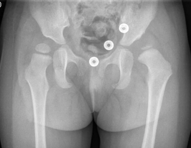
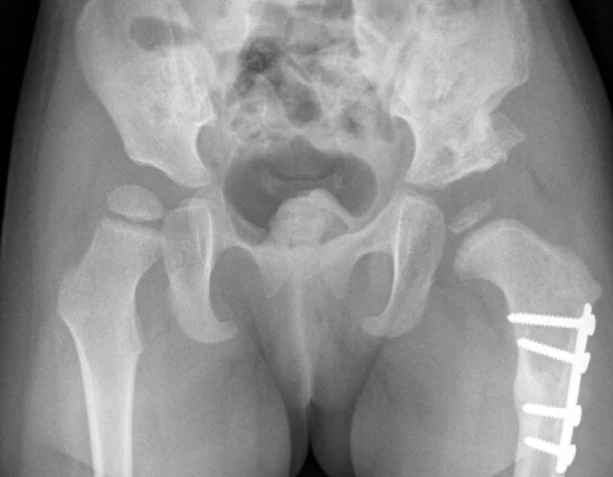
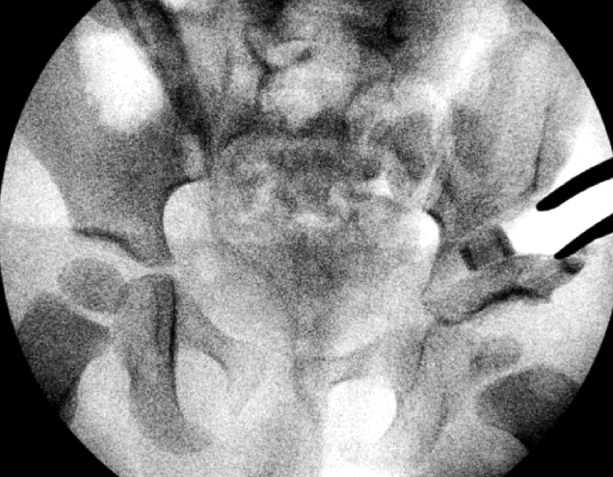
Clinical Case A : 3 year old boy with residual hip dysplasia after previous open reduction
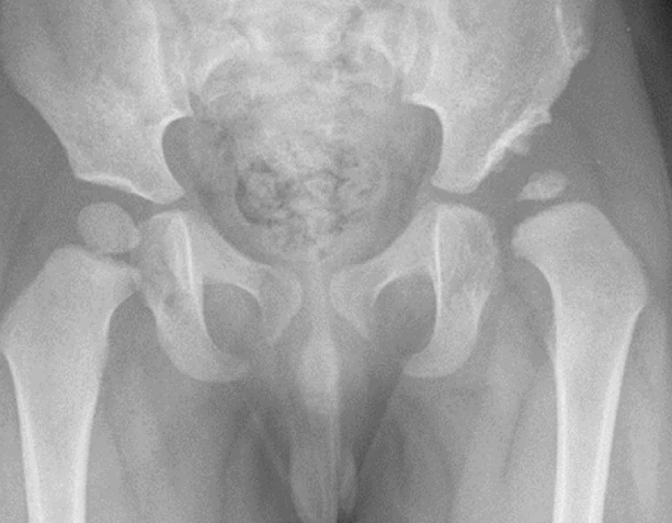
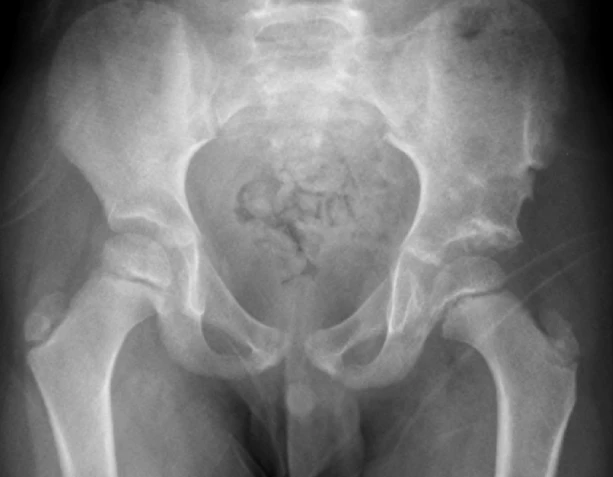
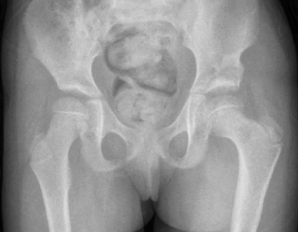
Clinical Case B :18 month old girl with subluxation after open reduction