Working time : Mon – Fri: 09:00AM – 5:00PM

Give Us a Call : + 0800 24 66 721

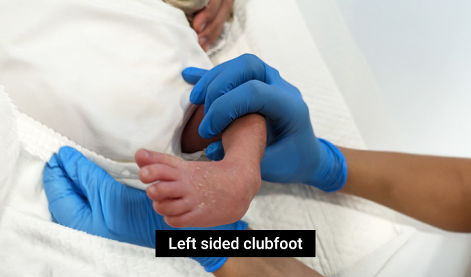
What is a clubfoot ?

A clubfoot is one of the commonest congenital lower limb problems that occur in children. The term congenital implies that it is present at birth. It may affect one or both feet and affects 1 in 1000 births. The clubfoot has a characteristic shape. Viewed from below the sole of the foot looks bean shaped with a curved outer border. The sole of the foot points inwards and the normal arch is accentuated. The important finding in a “true” clubfoot is that the foot is a little stiff. If you try to straighten a clubfoot, you will usually feel some resistance. Some clubfeet may have some flexibility. Other clubfeet can be very stiff. However, they all demonstrate some resistance in trying to push the foot straight.

Start your child’s journey to healthy feet today – learn more about the Ponseti method now!

What is a positional/postural clubfoot?

Many babies are born with feet that look like clubfeet with the feet curled in. This happens because the feet have been curled up in this position in the uterus during the latter weeks of pregnancy. However, these are not “true” clubfeet because the foot can easily be stretched with minimal effort. Simple handling can make the foot straight and lift the foot up towards the shin. This is a positional or postural clubfoot. It is a flexible deformity that can easily be stretched straight. It responds to simple stretching exercises which can be done by the parents and improves within the first few weeks of life. The true clubfoot does not easily stretch out and requires treatment to make the foot straight.

It is important to ensure that a positional clubfoot which will get better on its own is not subjected to lengthy and intrusive treatment which is not required. Therefore it is always best to see a Consultant Pediatric Orthopedic Surgeon who is experienced in recognizing and treating clubfoot.

What causes clubfoot?

The exact cause of clubfoot has yet to be determined. This is why it is termed “idiopathic” clubfoot. It can be seen more commonly in certain families due to a genetic tendency. However, in the vast majority of cases there is no family history of clubfoot. It is important to distinguish the idiopathic clubfoot from similar deformities which arise from other conditions. These include nerve and muscle conditions such as spina bifida, conditions affecting the brain such as cerebral palsy and syndromes affecting muscles and nerves such as arthrogryposis. These types of clubfoot may not necessarily be present at birth but develop later. They often result in stiffer deformities which can be more resistant to conventional treatment.

Can clubfoot be diagnosed before birth?

Although most clubfoot are first detected after birth, in some cases they may be diagnosed on the antenatal ultrasound scan during pregnancy. Often this can create a lot of anxiety in parents leading up to the birth of their child. It is important to offer pre-natal counselling to expectant mothers where an US diagnosis of probable clubfoot has been made. Discussion and reassurance on the diagnosis and response to treatment has a very positive impact on allaying parental concerns in the run up to the birth of their child. Dr Qureshi is happy to see expectant parents who have been given an antenatal diagnosis of possible clubfoot for counselling, guidance and advice.

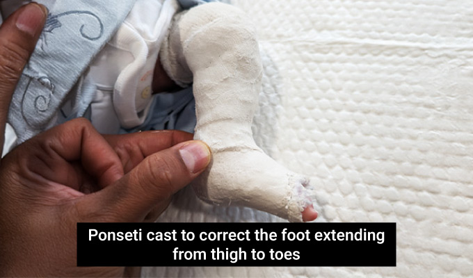
How do we treat clubfoot?

The internationally recognized and agreed treatment protocol for idiopathic clubfoot is Ponseti treatment. The technique involves serial casting with a small clinic procedure near the end. A defined sequence of weekly manipulative stretches and cast applications slowly brings the foot into a normal position.
Each week the foot is manipulated and a plaster cast is applied from the toes to the thigh to hold the foot in the newly achieved position. After one week the cast is removed and the foot is manipulated to stretch it into the next stage of the improvement. Once again a new cast is applied in this improved position. The casts are changed on a weekly basis until the foot is straight and pointing outwards.
The final stage of the foot correction is correcting the equinus (the foot pointing down). Unfortunately, the foot can not be brought up with stretching alone. It requires the achilles tendon to be cut. This is usually done as a clinic procedure. Local anaesthetic cream (numbing cream) is applied to the skin and a pea sized amount of local anesthetic is injected around the tendon. Using a fine scalpel, the tendon is cut through a very small incision. It is obvious when the tendon has been completely divided as the foot can be brought up easily. The final cast is applied in this corrected position with the foot pointing upwards and outwards. The final cast remains for 3 weeks to enable the tendon to heal in an elongated position

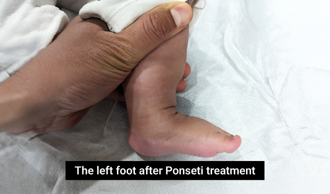
After Ponseti casting and tenotomy, is the clubfoot cured?

After completion of casting and tenotomy, the feet need to be braced during development to stop the deformity returning. The bracing consists of application of boots connected to a bar. This holds the feet in a nicely corrected position. The boots and bar are worn 23 hours a day for 3 months following final cast removal. After 3 months, they should be applied 12 hours a day (typically when the child is asleep) up to the age of 4 to 5 years.
Clubfoot is a developmental condition. If the bracing protocol is not strictly adhered to, the deformities will return as the foot grows. Bracing during the crucial years of early development is vital to maintain the correction and avoid the need for repeat casting. Clubfoot treatment should be viewed as a journey and not a discrete event. Children with clubfeet should be followed up with structured surveillance up to 10 -12 years of age to ensure that the feet are comfortable with good function.

Schedule your child’s orthopedic consultation in Dubai today! 

Dr. Assad Qureshi

Dr. Assad Qureshi is a highly experienced Pediatric Orthopedic Surgeon specializing in musculoskeletal disorders in children. With a focus on early diagnosis and advanced surgical techniques, he is committed to restoring function and improving the quality of life for his pediatric patients.